Translate this page into:
Chronic recalcitrant paronychia in a farmer
*Corresponding author: Eckart Haneke, Department of Dermatology, Inselspital, University of Bern, Bern, Switzerland. haneke@gmx.net
-
Received: ,
Accepted: ,
How to cite this article: Haneke E, Gabutti MP, Pelzer C, Iorizzo M. Chronic recalcitrant paronychia in a farmer. J Onychol Nail Surg. 2024;1:41-4. doi: 10.25259/JONS_13_2024
Abstract
Chronic paronychia is a common condition mainly afflicting middle-aged women. It is usually due to long-standing and repeated irritation and contact with moisture. We observed a male farmer with chronic paronychia. Cow hair was found under the proximal nail fold of the right middle finger. Bacterial culture revealed Mycobacterium elephantis in addition to mixed aerobic and anaerobic bacteria. Avulsion of the radial nail strip and phenolisation of the matrix horn led to a rapid and sustained resolution.
Keywords
Chronic paronychia
Cow hair
Mycobacterium elephantis
Treatment
Foreign body reaction
INTRODUCTION
Paronychia is an alteration of the nail folds and immediate surrounding skin of the nail unit. It may be acute and caused by trauma or infection, or chronic and due to a variety of infections and repeated irritation. Usually, paronychia lasting longer than 6 weeks is categorised as chronic paronychia. Most cases are seen involving the fingers of women aged 30–60 years. Its aetiology is often not entirely clear. It is assumed that repeated trauma due to long-term contact with water and chemical solutions, allergic reactions, and mechanical manipulation may lead to injury to the cuticle and loss of its sealing function for the nail pocket. This plays an important role in the development of chronic paronychia. Secondary microbial contamination or even infection is common. The treatment is always challenging as the condition is often self-aggravating.
CASE REPORT
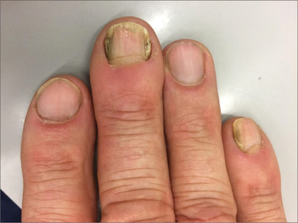
A 63-year-old male Swiss farmer was referred with recalcitrant greenish discolouration of the radial side of the right little fingernail and both sides of the right middle fingernail [Figure 1]. Treatment with soaks of diluted white vinegar and octenidine hydrochloride 2% solution, several times a day was recommended. While the little fingernail and the ulnar side of the middle fingernail healed within several weeks, the radial side of the nail improved only slightly. Infact, over the course of 8 months, it became worse and developed painful swelling with reddening of the radial quarter of the proximal nail fold. The fold appeared retracted and lacked the cuticle laterally. Its free margin was rounded in the radial quarter. The nail was dirty greenish in colour, and showed distal-lateral onycholysis [Figure 2]. Probing allowed the instrument to be inserted under the proximal nail fold virtually to the bottom of the culde-sac. However, over the following 8 weeks, the conditions worsened and became more painful [Figure 3].

- Paronychia of the right middle and fifth finger at first consultation.

- Paronychia of the radial half of the proximal nail fold of the right middle finger, ten months after the initial presentation.

- Paronychia of the radial half of the proximal nail fold of the right middle finger, one year after initial presentation.
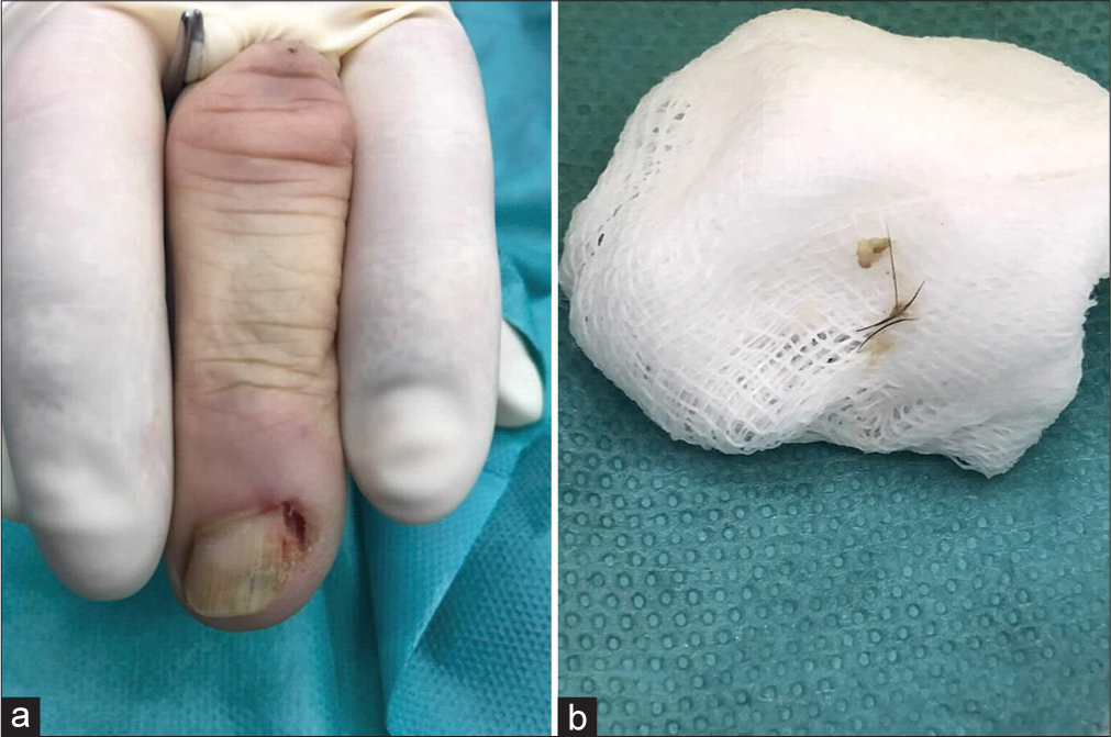
The patient was scheduled for surgical avulsion of the radial nail strip under block anaesthesia with 0.5% ropivacaine. After completion of the anaesthesia, the space under the proximal nail fold was cleaned, yielding characteristic cow hairs. A narrow lateral strip of the nail was cut longitudinally and avulsed [Figure 4]. The most lateral portion of the matrix was cautiously cauterised with 88% phenol, 3 cycles of 1 min each [Figure 4a]. Healing was uneventful and completed within 10 days [Figure 5]. The cuticle formed spontaneously again over 4 weeks. No specific antibiotic therapy was needed. Histopathology of the nail strip demonstrated plenty of bacteria on its undersurface, but periodic acid–Schiff stain did not show invading fungi. A bacterial and mycological swab of the affected area was taken. The bacterial culture yielded a mixed aerobic and anaerobic flora as well as Mycobacterium elephantis (100–200 colonies: ++) on selective culture media. The patient re-consulted 2 years after the surgery, reporting that he had been free from paronychia for approximately 18 months. However, the non-operated ulnar side of the right middle fingernail had now developed an insidious inflammation with thickening of the soft tissue and disappearance of the cuticle of the ulnar half of the proximal nail fold, roughly 6 months ago. As requested, another identical intervention was performed on the ulnar half. Healing was again uneventful. After 18 months, the right middle fingernail inflammation was completely resolved [Figure 6].

- (a) The affected radial nail plate margin has been avulsed. (b) Cow hairs extracted from under the proximal nail fold.

- Condition of the nail fold 24 hours after phenolisation.

- Outcome of surgery, three years after phenolisation.
DISCUSSION
Chronic paronychia is a relatively common condition affecting more women than men. It has been observed with many different occupations, particularly where prolonged contact with water or other fluids is associated with chronic repeated trauma.[1] The aetiology is often not entirely clear; however, wet work and manicures appear to play a major pathogenetic role. If these predisposing factors are not corrected, recurrence of paronychia is frequent. Once the cuticle has disappeared, the underlying nail detaches from the eponychium, and foreign bodies easily get trapped under the proximal nail fold. The patients are usually not able to identify as to what happened first, the loss of the cuticle or the inflammation. Wet work renders the skin soft, damages the cuticle, and allows foreign bodies to penetrate. Candida albicans was long held responsible for chronic paronychia, particularly in homemakers, chefs, butchers, fishmongers, and other similar wet-work professions. An immediate-type of contact allergic reaction to food ingredients has been shown to be a potential cause, and treatment with potent topical corticosteroids is recommended.[2,3] Chronic allergic contact dermatitis is also frequently seen and may be suspected on histopathology and proven by patch test.[4] Our patient had no history of contact allergy but carried out hard manual work as a farmer raising cattle, leading to chronic and repeated trauma.
The course of chronic paronychia is commonly prolonged, as was seen in our patient. Green nail discolouration suggests colonisation, or even infection, with Pseudomonas aeruginosa, which often responds to soaks with dilute vinegar, bleach, or antiseptic solutions, such as octenidine.[5] After an initial improvement, paronychia recurred on one side of the middle fingernail, even getting worse. The therapeutic problems were discussed with the patient who agreed for minor surgical intervention with avulsion of the lateral nail strip and phenolisation of the matrix horn. This led to an immediate improvement and sustained resolution. During the surgical procedure, cow hairs were retrieved from under the nail fold. This phenomenon is known as an occupational disease in hairdressers, with human hair shafts getting trapped under the proximal nail fold causing chronic paronychia[6,7], or piercing the skin (interdigital space, or under the nail) with resultant interdigital pilonidal sinus formation.[8-10] Cow hair is harder than human hair. It can be clinically differentiated from human hair by its thickness and tapered ends.[11]
The entrapment of foreign bodies such as (cow) hairs under the nail fold predisposes to a chronic foreign-body reaction that, over weeks and months, leads to a fibrotic thickening with rounding of the fold’s free margin.[12,13] This results in the loss of the cuticle and is a perfect example of a vicious cycle.[1] Such a reaction does not remain sterile; hence, the demonstration of bacteria and fungi, mainly Candida spp., is common. In our patient, both aerobic and anaerobic bacteria were cultured including P. aeruginosa. To the best of our knowledge, M. elephantis has never been isolated from chronic paronychia and was a surprise finding. This organism has most commonly been observed in humans with chronic lung conditions[14-18] and recently as a cutaneous infection with lymphocutaneous spread.[19] A few reports of this infection exist in cows, explaining its presence in our patient.[20-23] However, its pathogenetic role in our case remains doubtful as paronychia healed rapidly after matrix horn phenolisation. Subsequent cultures also failed to demonstrate the organism. This could be because phenol is a potent antiseptic agent, and the lateral horns (potentially contaminated with P. aeruginosa and M elephantis) were eliminated with phenolisation.
CONCLUSION
This case is reported to present recalcitrant chronic paronychia in a farmer due to cow hair and potentially M. elephantis, to raise awareness about the atypical features.
Authors’ contributions
EH has seen the patient, made the diagnosis, performed the biopsy, saw the histopathology, performed the treatment, drafted and wrote the manuscript. MPG has seen the patient, performed the biopsy, and the treatment, and read the manuscript. MI and CP proofread the manuscript and organised the manuscript.
Ethical approval
Institutional Review Board approval is not required.
Declaration of patient consent
The authors certify that they have obtained all appropriate patient consent.
Conflicts of interest
Dr. Matilde Iorizzo and Dr. Eckart Haneke are on the editorial board of the Journal.
Use of artificial intelligence (AI)-assisted technology for manuscript preparation
The authors confirm that there was no use of artificial intelligence (AI)-assisted technology for assisting in the writing or editing of the manuscript and no images were manipulated using AI.
Financial support and sponsorship
Nil.
References
- Acute and chronic paronychia revisited: A narrative review. J Cutan Aesthet Surg. 2022;15:1-16.
- [CrossRef] [PubMed] [Google Scholar]
- Role of foods in the pathogenesis of chronic paronychia. J Am Acad Dermatol. 1992;27:706-10.
- [CrossRef] [PubMed] [Google Scholar]
- Topical steroids versus systemic antifungals in the treatment of chronic paronychia: An open, randomized double-blind and double dummy study. J Am Acad Dermatol. 2002;47:73-6.
- [CrossRef] [PubMed] [Google Scholar]
- Histopathological analysis of chronic paronychia. Int J Dermatol. 2023;62:514-7.
- [CrossRef] [PubMed] [Google Scholar]
- Chronic paronychia in which hair was a foreign body. Int J Dermatol. 1975;14:661-3.
- [CrossRef] [PubMed] [Google Scholar]
- Chronic paronychia in a hairdresser. Occup Med (Lond). 2014;64:468-9.
- [CrossRef] [PubMed] [Google Scholar]
- Barber's hair sinus in a female hairdresser: Uncommon manifestation of an occupational dermatosis. J Eur Acad Dermatol Venereol. 2006;20:209-11.
- [CrossRef] [PubMed] [Google Scholar]
- Onycholysis associated with subungual manifestation of barber's hair sinus. Int J Dermatol. 2007;46(Suppl 3):48-9.
- [CrossRef] [PubMed] [Google Scholar]
- Barber's hair sinus in a female hairdresser: Uncommon manifestation of an occupational disease: A case report. Cases J. 2008;1:1-4.
- [CrossRef] [PubMed] [Google Scholar]
- Milker dermatoses. Milker's nodules with exanthem. Nail changes from cow hairs. Arch Dermatol Syphilis. 1932;164:603-9.
- [CrossRef] [Google Scholar]
- Phenotypic and molecular characterization of clinical isolates of Mycobacterium elephantis from human specimens. J Clin Microbiol. 2002;40:1230-6.
- [CrossRef] [PubMed] [Google Scholar]
- Recovery of Mycobacterium elephantis from sputum of a patient in Belgium. J Clin Microbiol. 2003;41:1344.
- [CrossRef] [PubMed] [Google Scholar]
- Mycobacterium elephantis Not an exceptional finding in clinical specimens. Eur J Clin Microbiol Infect Dis. 2003;22:427-30.
- [CrossRef] [PubMed] [Google Scholar]
- First report of isolation of Mycobacterium elephantis from bronchial lavage of a patient in Asia. JRSM Short Rep. 2011;2:26.
- [CrossRef] [PubMed] [Google Scholar]
- Draft genome sequence of Mycobacterium elephantis strain Lipa. Genome Announc. 2015;3:e00691-15.
- [CrossRef] [PubMed] [Google Scholar]
- Lymphocutaneous spread of Mycobacterium elephantis in an immunocompetent individual: A case report. SAGE Open Med Case Rep. 2021;9:2050313X211034913.
- [CrossRef] [PubMed] [Google Scholar]
- First cases of Mycobacterium elephantis in Zimbabwe revealed by 16s ribosequencing. Arch Clin Microbiol. 2015;6:10-3.
- [CrossRef] [PubMed] [Google Scholar]
- Molecular identification of Mycobacterium species of public health importance in cattle in Zimbabwe by 16S rRNA gene sequencing. Open Microbiol J. 2015;9:38-42.
- [CrossRef] [PubMed] [Google Scholar]
- First report in China on the identification and drug sensitivity of Mycobacterium elephantis isolated from the milk of a cow with mastitis. Biomed Environ Sci. 2017;30:501-7.
- [Google Scholar]
- Identification and drug susceptibility testing of Mycobacterium thermoresistibile and Mycobacterium elephantis isolated from a cow with mastitis. Zhonghua Liu Xing Bing Xue Za Zhi. 2018;39:669-72.
- [Google Scholar]


