

Translate this page into:
Is there a difference between habit tic and median canaliform nail dystrophy of Heller?
*Corresponding author: Eckart Haneke, Department of Dermatology, Inselspital, University of Bern, Bern, Switzerland. haneke@gmx.net
-
Received: ,
Accepted: ,
How to cite this article: Haneke E. Is there a difference between habit tic and median canaliform nail dystrophy of Heller? J Onychol Nail Surg. 2025;2:4-9. doi: 10.25259/JONS_8_2025
Abstract
Heller’s Median canaliform nail dystrophy (MCD) and habit tic deformity (HTD) are often confused with each other, although there are distinct differences between the two conditions. MCD is characterised by a median break in the nail and obliquely-proximally running furrows without a longitudinal depression. Signs of skin picking are lacking. HTD shows longitudinal shallow and wide depression with innumerable short transverse lines, due to repeated pressure on the nail matrix as a result of repeated pushing of the proximal nail fold’s free margin back.
Keywords
Body-focused repetitive behaviour
Differential diagnosis
Dystrophia mediana unguium canaliformis
Habit tic
Median canaliform nail dystrophy
Onychopapilloma
Solenonychia

Autoaggressive behaviours targeting the nails are a well-defined group of conditions in the large field of body-focused repetitive behaviours (BFRB). They are usually divided into habit tic deformity (HTD), nail chewing (onychophagia), the group of onychotillomania – onychoteiromania – onychotemnomania, and overzealous manicure.
HTD often includes picking of the perionychial skin.[1] The characteristic habit-tic involves a persistent pushing back of the proximal nail fold by pressing the nail of another finger (usually the thumbnail of the opposite hand) onto the proximal nail plate, and then moving it forcefully proximally to push the cuticle back. This leads to a series of changes in the nail and the proximal nail fold [Figure 1]. There is a loss of the sharp angle of the proximal nail fold’s free margin, with the disappearance of the cuticle and apparent thickening of the nail fold. This results in a detachment of the nail fold’s free margin from the underlying nail plate. Subsequent changes include an excessively long and large lunula (mega lunula) and a thumbnail that appears markedly longer than wide [Figure 2]. Serial median transverse furrows appear over the nail plate, with a central depression, giving the nail surface the appearance of a washboard. The medial depression and the shallow longitudinal furrows occur due to chronically repeated trauma on the nail plate, which is still soft when it emerges from under the proximal nail fold.

- Schematic illustration of (a) a normal nail plate surface for reference; and (b) the appearance in cases with habit tic deformity; (c) median canaliform dystrophy; and (d) solenychia.
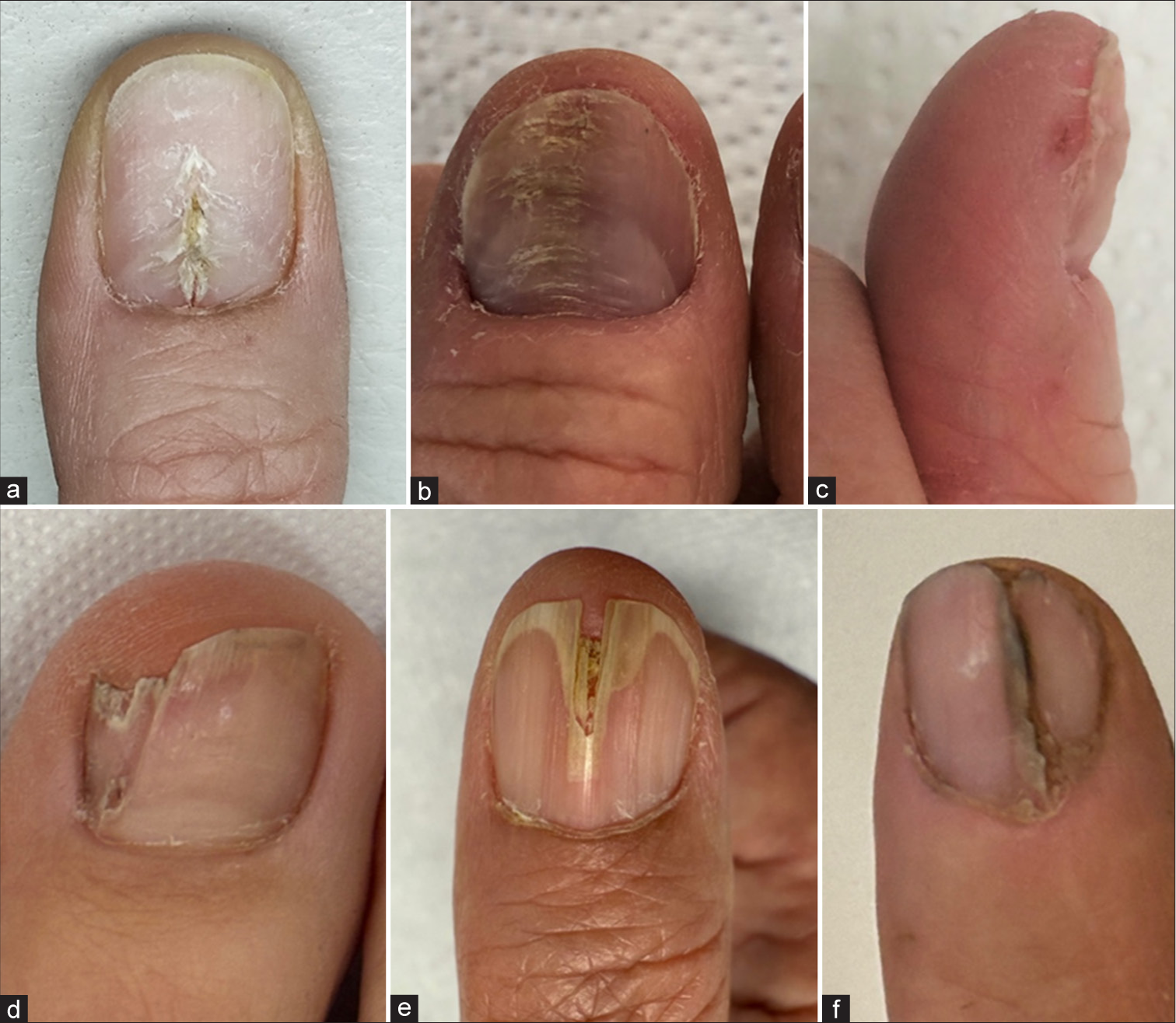
- Nails with longitudinal median or paramedian dystrophy. (a) Thumbnail showing median canaliform dystrophy demonstrating a median split in the proximal part of the nail plate with oblique furrows giving the aspect of a fir tree. (b) Another thumbnail showing habit-tic deformity with enormous enlargement of the lunula, loss of the cuticle and wash-board appearance due to serial short transverse furrows and signs of skin picking. (c) Thumbnail with habit-tic deformity seen from the side exhibits a deep depression in the proximal nail region, thickening of the free margin of the proximal nail fold, loss of cuticle and severe perionychotillomania. (d) Great toenail showing a paramedian intraungual fibrokeratoma causing ‘solenonychia’. (e) Thumbnail showing onychopapilloma in median location. (f) Traumatic nail split.
These usually cover at least one-third of the nail’s width. The proximal nail plate just in front of the nail fold may appear sinking inwards, accentuating the transition from the nail fold, which appears thickened. In advanced cases, the proximal nail fold’s free margin may be completely pushed back to the depth of the nail pocket, completely obliterating it. This results in roughness involving the medial one-third to one-half of the nail plate because the most apical matrix, responsible for producing the shiny superficial nail plate, is damaged. Intense manipulation may even lead to superficial lamellar nail plate defects, similar to elkonyxis. This HTD is often associated with skin picking, also known as perionychotillomania.
In contrast, Heller’s median canaliform (thumb) nail dystrophy (MCD) is characterised by a single longitudinal fissure or break in the thumbnail, with no depression in the plate. It starts at the cuticle, and slowly grows distally. Obliquely oriented, proximally directed furrows originate from the median fissure, giving the nail the appearance of a “fir-tree” [Figure 1]. This longitudinal crack often does not reach the free margin of the nail plate. In MCD, transverse furrows are not a characteristic feature, the nail is not longitudinally depressed, and the lunula may or may not be increased in size. Obvious pushing back or thickening of the free margin of the proximal nail fold is not apparent. In most cases, the cuticle is intact [Figure 2].
Signs of perionychotillomania are usually lacking, although some cracking of the cuticle may be seen.[2] Thus, these two conditions can be distinguished largely based on their clear morphological differences.[3] Unfortunately, this is often not the case as depicted by the two manuscripts submitted to this issue of JONS.[4,5] A thorough literature search of these two conditions showed that the confusion has been prevalent in a number of other publications [Table 1].[2,6-43] All the references were not available as full text[44-57], also the images were not clear.[58]
| Authors and year | Journal | Claimed diagnosis | Correct diagnosis | Additional findings |
|---|---|---|---|---|
| Heller, 1928[2] | Dermatol Z 1928:416 | MCD | MCD | First description |
| Kallos, 1948[6] | Dermatologica 96:432 | MCD | MCD | Healed with X-ray |
| Robinson and Weidman, 1948[7] | Arch Dermatol 57:328 | MCD | Possibly Onycho-papilloma | - |
| Leclercq, 1964[8] | Bull Soc Fr Dermatol Syphiligr 71:655-8 | MCD | **Full text not found, Article in French | - |
| Hoffmann-Martinot, 1964[9] | Bull Soc Fr Dermatol Syphiligr 71:759 | MCD | Myxoid pseudocyst | - |
| Sutton, 1965[10] | South Med J 58:1143 | Solen-onychia=MCD | Intraungual fibrokeratoma | Terminology discussed but incorrect |
| van Dijk, 1978[11] | Dermatologica 156:358 | MCD | **Full text not found | - |
| Guerra et al., 1988[12] | G Ital Dermatol Venereol 123:555 | MCD | **Full text not found | Caused by trauma |
| Braun-Falco et al., Springer 1991: 790[13] | Dermatology 1991 Figure 32: 12 Figure 32: 13 | MCD MCD | HTD MCND | One correct and one wrong diagnosis in the same chapter |
| Bottomley and Cunliffe, 1992[14] | Br J Dermatol 127:447 | MCD | MCD | Associated with isotretinoin |
| Griego et al., 1995[15] | Int J Dermatol 34:799 | MCD HTD | MCD (Rt) HTD (Lt) | Patient had MCD on the right thumb and HTD on the left thumb |
| Dharmagunawardena and Charles-Holmes, 1997[16] | Br J Dermatol. 137:658 | MCD | MCD | Following isotretinoin |
| Sweeney et al., 2005[17] | Cutis. 2005;75 (3):161 | Familial MCD | HTD | Familial |
| Gloster and Kindred, 2005[18] | J Am Acad Dermatol 53:543 | MCD | HTD | Improved with multivitamins |
| Verma, 2008[19] | Indian J Dermatol Venereol 74:257 | Nail split due to glomus tumour | same | No MCD as claimed |
| Wu et al., 2009[20] | J Eur Acad Dermatol Venereo. 23:1102 | MCD | MCD (Rt) HTD (Lt) | Patient had MCD on the right and HTD on the left thumb |
| Olszewska et al., 2009[21] | Am J Clin Dermatol; 10:193 | MCD (PDA nail) | HTD | Cleared after repeated pressure was stopped |
| Kim et al., 2010[22] | J Dermatol 37:573 | MCD | HTD | Treatment with Tacrolimus |
| Latham and Langley, 2013[23] | Can Fam Physician. 2013;59:511 | MCD | MCD | Differential diagnosis of onychomycosis |
| Avhad and Ghuge 2013[24] | Ind J Ped 50:1073 | MCD | HTD | - |
| Borges-Costa et al., 2013[25] | Int J Dermatol 52:1581 | MCD | Median ridging | Appeared during ritonavir therapy |
| Winther and Bygum, 2014[26] | Acta Derm Venereol 94:719 | MCD | Mixed features | Appeared during alitretinoin treatment |
| Schmutz and Tréchot, 2014[27] | Ann Dermatol Venereol 141:485 | MCD | **Full text not found | Associated with ritonavir |
| de Roos, 2015[28] | Ned Tijdschr Geneeskd 159:A8471 | MCD | MCD | - |
| Kota et al., 2016[29] | Indian J Dermatol 61:120 | MCD | HTD | - |
| Alli and Dogan, 2016[30] | Cutan. Ocul Toxicol 35:85 | MCD | MCD plus elkonyxis | Due to short-term isotretinoin |
| Pathania, 2016[31] | Med J Armed Forces India 72:178 | MCD | HTD | Tacrolimus and fluoxetine |
| Choi et al., 2017[32] | J Cosmet Laser Ther 19:225 | MCD | MCD | Long-pulse Nd-YAG laser treatment |
| Damevska et al., 2017[33] | Pediatr Dermatol 34:726 | MCD | HTD-like | Developed after cryotherapy |
| Jain et al., 2019[34] | Ind J Ped Dermatol 21:53 | MCD | HTD | |
| Wang et al., 2020[35] | Australas J Dermatol. 61:e100 | MCD plus HTD | HTD | Atopic dermatitis |
| Giura et al., 2020[36] | Clin Exp Dermatol 45:601 | MCD | MCD | Atopic dermatitis present. MCD cleared with dupilumab |
| Khodaee et al., 2020[37] | CMAJ 192:E1810 | MCD | HTD | |
| Quan and Johnson, 2022[38] (Commented by Sloan, 2023)[39] | JAAD Case Rep 29:70 (Comment JAAD Case Rep 29:325) | MCD | MCD (Comment: solenonychia) | Treatment topical tazarotene. (Comment wrong) |
| Wilson et al., 2023[40] | Pediatr Dermatol 40:511 | MCD | MCD | 2-y-o boy, treated marigold cream |
| Pinto et al., 2023[41] | BMJ Case Rep 16 (7):e257251 | MCD | HTD | Body-focused repetitive behaviour |
| Pauliņa and Balcere, 2023[42] | Dermatol Pract Concept. 13:e2023184 | MCD | MCD | Associated with retinoid therapy |
| El Fatoiki et al.[43] | Skin Appendage Disord 10:236 | MCD | HTD -like | Professional trauma |
MCD: Median canaliform nail dystrophy; HTD: Habit-Tic deformity **Not all references were available as full text.
On searching ‘median canaliform nail dystrophy’ on Pubmed, the term solenonychia also appears. However, on further search, it was found to be coined for a nail disorder characterised by a paramedian canal in the plate of one nail.[10] The figures in this publication show a narrow, sharply demarcated canal in the big toenail. Evaluating the photographs revealed an intraungual fibrokeratoma, a diagnosis not related to Heller’s MCD or HTD. The author in this report had discussed the term MCDN, elaborating that his term “solenonychia” is more representative. Unfortunately, the correct diagnosis seems to have been missed.
A regular longitudinal depression in the nail may also be seen as a result of pressure on the nail matrix by myxoid pseudocysts. In type B lesions, a rupture of the pseudocyst into the nail pocket is frequent, leading to a temporary decrease in the pressure on the matrix. This results in interruptions in the longitudinal depression, intermittently. However, observations show this type of midline depression to be completely different from the transverse furrows of the HTD. Distal nail splits are also commonly seen in cases of onychopapilloma. However, these are also different from the proximally beginning crack of Heller’s MCD. A variety of subungual tumours may also cause a longitudinal split in the nail plate due to the volume of the neoplasm. Similarly, longitudinal scars as well as cicatricial pterygium may also occasionally cause a (para)median split in the nail plate; however, the characteristic oblique furrows are lacking.
Both HTD and the MCD are caused by repeated trauma. This is more obvious in cases with HTD, but less so in Heller’s MCD. Virtually all patients with BFRB tend to reject the autoaggressive etiology of their complaints; however, HTD patients may eventually acknowledge this after counseling. The type of mechanical trauma in MCD is much more difficult to ascertain. Most patients are truly not aware of a specific behaviour leading to this outcome. Consistent protection of the nail region with a foam tube, an occlusive dressing such as skin-colored suture strips, or an artificial nail, invariably leads to improvement and, finally, the disappearance of the median nail split. Sometimes, even therapies not specifically indicated, such as antivirals or retinoids, have been reported to produce improvement, as the patient is made aware of the condition, and takes care not to traumatise the thumbnail [Table 1]. In contrast, stopping the habit-tic is more difficult. The patient may not be aware of the habit and it requires consistent, empathetic explanation to recognise this behaviour, without causing offense. Many different measures have been proposed. These include simple mechanical devices, behaviour-altering psychotherapy, and even psychopharmacologic drugs. Recently, N-acetyl cysteine has been used for obsessive-compulsive disorders with some effect. The usual dose for adults is 1800–2400 mg per day.[59]
To conclude, habit-tic deformity and median canaliform dystrophy are distinct entities, which can be recognized clinically, based on subtle differences. This recognition aids appropriate management of these cases.
Ethical approval:
Institutional Review Board approval is not required.
Declaration of patient consent:
Patient’s consent was not required as there are no patients in this study.
Conflicts of interest:
Dr. Eckart Haneke is on the editorial board of the Journal.
Use of artificial intelligence (AI)-assisted technology for manuscript preparation:
The authors confirm that there was no use of artificial intelligence (AI)-assisted technology for assisting in the writing or editing of the manuscript and no images were manipulated using AI.
Financial support and sponsorship: Nil.
References
- Zur Kasuistik seltener nagelkrankheiten: Dystrophia unguium mediana canaliformis. Dermatol Zschr. 1928;51:416-9.
- [CrossRef] [Google Scholar]
- Self-induced nail disorders In: Rubin AI, Daniel CR 3rd, Lipner SR, Haneke E, Jellinek N, Scher RK, eds. Scher and Daniel's nails (5th ed). Berlin: Springer International Springer Nature; 2025.
- [Google Scholar]
- ‘Washboard nails’ secondary to habit-tic deformity versus median canaliform dystrophy of Heller: The morpho-diagnostic conundrum. J Onychol Nail Surg. 2025;2:54-6.
- [Google Scholar]
- Habit-tic deformity or median canaliform dystrophy-at a crossroad. J Onychol Nail Surg. 2025;2:57-9.
- [Google Scholar]
- Dystrophia unguium mediana canaliformis (Heller) Dermatologica. 1948;96:432-3.
- [CrossRef] [PubMed] [Google Scholar]
- Dystrophia unguium mediana canaliformis. Arch Derm Syphilol. 1948;57:328-31.
- [CrossRef] [PubMed] [Google Scholar]
- Naevus striatus symetricus unguis, dystrophie médiane canaliforme de Heller ou dystrophie unguéale médiane en chevrons. Etude nosologique à propos de deux cas [nevus striatus unguis, median canaliform dystrophy of Heller or median onychoid dystrophy in chevrons. Nosologic study apropos of 2 cases] Bull Soc Fr Dermatol Syphiligr. 1964;71:655-8.
- [Google Scholar]
- Deux cas de dystrophie unguéale canaliculaire médiane par kyste de la face dorsale de la dernière phalange [2 cases of median canalicular ungueal dystrophy due to cyst of the dorsal surface of the last phalanx] Bull Soc Fr Dermatol Syphiligr. 1964;71:759.
- [Google Scholar]
- Solenonychia: Canaliform dystrophy of the nails. South Med J. 1965;58:1143-6.
- [CrossRef] [Google Scholar]
- Dystrophia unguium mediana canaliformis. Dermatologica. 1978;156:358-66.
- [CrossRef] [PubMed] [Google Scholar]
- Distrofia ungueale mediana traumatica [Median ungual dystrophy caused by trauma] G Ital Dermatol Venereol. 1988;123:555-6.
- [Google Scholar]
- Median nail dystrophy associated with isotretinoin therapy. Br J Dermatol. 1992;127:447-8.
- [CrossRef] [PubMed] [Google Scholar]
- Median nail dystrophy and habit tic deformity: Are they different forms of the same disorder? Int J Dermatol. 1995;34:799-800.
- [CrossRef] [PubMed] [Google Scholar]
- Median canaliform dystrophy following isotretinoin therapy. Br J Dermatol. 1997;137:658-9.
- [CrossRef] [PubMed] [Google Scholar]
- Habit-tic-like and median nail-like dystrophies treated with multivitamins. J Am Acad Dermatol. 2005;53:543-4.
- [CrossRef] [PubMed] [Google Scholar]
- Glomus tumor-induced longitudinal splitting of nail mimicking median canaliform dystrophy. Indian J Dermatol Venereol Leprol. 2008;74:257-9.
- [CrossRef] [PubMed] [Google Scholar]
- Median canaliform dystrophy of Heller with associated swan neck deformity. J Eur Acad Dermatol Venereol. 2009;23:1102-3.
- [CrossRef] [PubMed] [Google Scholar]
- Treatment of median canaliform nail dystrophy with topical 0.1% tacrolimus ointment. J Dermatol. 2010;37:573-4.
- [CrossRef] [PubMed] [Google Scholar]
- Median nail dystrophy associated with ritonavir. Int J Dermatol. 2013;52(12):1581-2.
- [CrossRef] [PubMed] [Google Scholar]
- Can median nail dystrophy be an adverse effect of alitretinoin treatment? Acta Derm Venereol. 2014;94:719-20.
- [CrossRef] [PubMed] [Google Scholar]
- Dystrophie unguéale médiane et ritonavir [Median nail dystrophy and ritonavir] Ann Dermatol Venereol. 2014;141:485-6.
- [CrossRef] [PubMed] [Google Scholar]
- Een man met nagelafwijkingen aan zijn duimen [A man with thumb nail abnormalities] Ned Tijdschr Geneeskd. 2015;159:A8471.
- [Google Scholar]
- Median nail dystrophy involving the thumb nail. Indian J Dermatol. 2016;61:120.
- [CrossRef] [PubMed] [Google Scholar]
- Short-term isotretinoin-induced elkonyxis and median nail dystrophy. Cutan Ocul Toxicol. 2016;35:85-6.
- [CrossRef] [PubMed] [Google Scholar]
- Median canaliform dystrophy of Heller occurring on thumb and great toe nails. Med J Armed Forces India. 2016;72:178-9.
- [CrossRef] [PubMed] [Google Scholar]
- Median canaliform nail dystrophy treated with a 1064-nm quasi-long pulsed Nd:YAG laser. J Cosmet Laser Ther. 2017;19:225-6.
- [CrossRef] [PubMed] [Google Scholar]
- Median canaliform dystrophy of Heller after cryotherapy. Pediatr Dermatol. 2017;34:726-7.
- [CrossRef] [PubMed] [Google Scholar]
- Median canaliform dystrophy of thumb and great toe nails in an 8 years old boy. Indian J Paediatr Dermatol. 2019;21:53-5.
- [CrossRef] [Google Scholar]
- Coexisting median canaliform nail dystrophy and habit-tic deformity in a patient with atopic dermatitis. Australas J Dermatol. 2020;61:e100-1.
- [CrossRef] [Google Scholar]
- Median canaliform nail dystrophy of Heller in a patient with atopic dermatitis: “Miraculous” healing with dupilumab. Clin Exp Dermatol. 2020;45:601-2.
- [CrossRef] [PubMed] [Google Scholar]
- Successful treatment of median canaliform nail dystrophy with topical tazarotene foam. JAAD Case Rep. 2022;29:70-1.
- [CrossRef] [PubMed] [Google Scholar]
- This month in JAAD case reports: February 2023 treatment option for solenonychia. J Am Acad Dermatol. 2023;88:325.
- [CrossRef] [Google Scholar]
- Median canaliform nail dystrophy in a 2-year-old boy: Case report and review of the literature. Ped Dermatol. 2023;40:511-8.
- [CrossRef] [PubMed] [Google Scholar]
- Median canaliform dystrophy of Heller presenting as a body-focused repetitive behaviour. BMJ Case Rep. 2023;16:e257251.
- [CrossRef] [PubMed] [Google Scholar]
- Median nail canaliform dystrophy in association with retinoid therapy. Dermatol Pract Concept. 2023;13:e2023184.
- [CrossRef] [PubMed] [Google Scholar]
- Median canaliform dystrophy of Heller in a carpet weaver: A new risk factor?-A case report. Skin Appendage Disord. 2024;10:236-8.
- [CrossRef] [PubMed] [Google Scholar]
- Ein fall von Dystrophia unguium mediana canaliformis Heller. Dermatol Zschr. 1928;54:96-8.
- [CrossRef] [Google Scholar]
- Dystrophia unguium mediana canaliformis. Arch Derm Syphilol. 1944;50:267-8.
- [CrossRef] [Google Scholar]
- Dystrophia unguium mediana canaliformis. AMA Arch Derm Syphilol. 1951;64:61-2.
- [CrossRef] [PubMed] [Google Scholar]
- Dystrophia unguium mediana canalformis periodica. Ann Dermatol Syphiligr (Paris). 1957;84:670-2.
- [Google Scholar]
- Zur klinik und pathogenese der dystrophia unguium mediana canaliformis (Heller) [Clinical picture and pathogenesis of dystrophia unguium mediana canaliformis (Heller)] Dermatol Wochenschr. 1958;137:380-6.
- [Google Scholar]
- Data on Heller's disease (dystrophia unguium mediana canaliformis)] Börgyógy Venerol Sz. 1960;36:159-63.
- [Google Scholar]
- Dystrophia unguium mediana canaliformis di Heller [Heller's dystrophia unguium mediana canaliformis] Minerva Dermatol. 1965;40:303-4.
- [Google Scholar]
- Etiologii dystrophia unguium mediana canaliformis Heller [On the etiology of dystrophia unguium mediana canaliformis Heller] Cesk Dermatol. 1966;41:164-7.
- [Google Scholar]
- Dystrophia unguis mediana canaliformis. Acta Dermatovener. 1971;51:315-17.
- [CrossRef] [Google Scholar]
- Correspondence: Median canaliform dystrophy of Heller induced by isotretinoin. J Dermatol Treat. 2000;11:135.
- [CrossRef] [Google Scholar]
- Dystrophia mediana canaliformis affecting all ten finger nails. Indian J Dermatol Venereol Leprol. 1995;61:58.
- [Google Scholar]
- Acetylcysteine for treatment of trichotillomania, excoriation disorder, onychophagia, and onychotillomania: An updated literature review. Int J Environ Res Public Health. 2022;19:6370.
- [CrossRef] [PubMed] [Google Scholar]


